Wednesday, March 27, 2013

Day 22: Stumbling upon Interesting Correlations

Taken from article.
Last night, while stumbling upon different science-related web pages through StumbleUpon, I found an article related to neuroscience! Tada! A Little Juice to the Brain Eases Depression. As I have discussed in a previous article, TMS is being used to treat depression and other mood disorders. This recently-published article talks not of TMS, but of transcranial direct current stimulation, or tDCS. What a familiar acronym! Where have I seen this before? Why, in one of my previous blog posts: "Day 12: Defense against the Dark rTMS." In my blog post, I read about how tDCS is used to negate the negative effects of rTMS.

In the article that I "stumbled upon," the experiment compared the affects of antidepressants vs. tDCS and found that tDCS was significantly better at treating depression than antidepressants. But the two together was found to have worked the fastest in treatment and was said to have had significant improvement by week 2.

The article I'm reading!
So now, I'm reading an article for Journal Club even though I will be absent Friday to visit the venue for Prom and to go to church for Good Friday! Anyway, the article is titled "Differential Modulation of Motor Cortical Plasticity and Excitability in Early and Late Phases of Human Motor Learning." I'll discuss more about it once I finish reading it.

For clarification: If you were wondering why the number of "Day"s is so small even though I've been at this for almost 2 months, the Day number is actually a day I'm in the lab. So, I've been in the lab for 22 individual days. I've been consistent, arriving here at ASU 3 days a week at 8am and starting work at 9.

References:
  1. Phend, C. "A Little Juice to the Brain Eases Depression." MedPage Today. 26 Feb 2013. http://www.medpagetoday.com/psychiatry/depression/37226?rXFb&xid=su_&hr=su&utm_source=stumbleupon&utm_medium=cpc&utm_campaign=psyc&rQZb/ (accessed March 25, 2010)

Tuesday, March 26, 2013

Day 21: Planning Data

Not much has been happening recently... But I have been making progress towards my final product.
click to enlarge

Today, there are high school students all around the lab taking a tour, so we're not doing any experiments or anything. I took this free day to import all the planning data for my project into my computer. It took me less than an hour to run the code for 7 subjects (6 blocks each) using the software Spike. We use Spike to view the muscle activity as MEP values and to create Excel spreadsheets with all the data (as shown to the right). Each column represents a different variable that will become apparent once the data goes through MatLab. Columns C, D, E, and F represent the MEP values for the muscles FDI (index finger), APB (thumb), ADM (pinky), and FCR (forearm), respectively. 

Looking through some data, I've seen a trend suggesting that positioning the fingers (P) requires surprisingly more excitability then positioning + applying a force (P+F). I originally expected to find that there wouldn't be a difference in excitability between the two because the movement for each action involves the same muscles. What I did not take into account is that the two actions are controlled by different parts of the brain. But in reality, excitability of P is greater than that of P+F. This is due to the inhibition of force application. During planning, the action of force application is inhibited because the action is more intense than just positioning the fingers. 

Something important to always keep in mind when reading this blog: This entire project is based on planning dexterous movements, which is why I imported all the planning data. Planning is the period after the cue (P or P+F) is given but before the movement is actually done. Related experiments that also use TMS focus on memory, learning, consciousness, and emotion (click here to learn more).

Thanks for reading my blog!

Saturday, March 23, 2013

Day 19: Another Day, Another Article

It was Friday yesterday which is Journal Club day! The article we read this week was titled "Action-blindsight in healthy subjects after transcranial magnetic stimulation." For those of you who don't know, blindsight is a condition caused by a lesion in the primary visual cortex, so it makes people blind in one side.

For methods, the experimenters used 11 subjects. The subjects sat in front of 3 buttons: Left button (LB), middle button (MB), and right button (RB). A subject was cued to move his or her hand to MB, and sometimes, a second cue would show up to correct them to either go left or right, which was chosen at random. The TMS had to be pulsed at a specific time period so that the subject could exhibit blindsight behavior. The TMS pulse caused the correction speed to decrease, which suggests the existence of an efference copy. This is a weird concept--a theoretical construct. Efference copy allows people to predict the effect of their movements. It enables a person to know the difference between actual movement and desired movement while the person moves. The action potentials for the movement don't have to go through an entire neural pathway, so it's like a shortcut, which is why in the experiment, the time taken for the subject to realize he or she have to correct his or her hand direction usually was lower than the amount of time the subject took to process the movement towards MB.
  • Fun fact: Have you ever tried tickling yourself? Most people are incapable of tickling themselves. It's a strange phenomenon caused by efference copy.
The study showed that TMS blocks conscious perception of the object placed on the blind side. Subjects of the trial claimed that they could perceive color spheres. But fortunately, TMS only INDUCED the blindsight behavior, so blind sight was not caused by injury or trauma. This reminds me of a kind of stimulation I've mentioned in a previous post: cTBS, a stimulation capable of creating virtual lesions. Crazy stuff, right?

Until my next adventure!

References:
  1. Christensen, M.S.; Kirstiansen, L.; Rowe, J.B.; Nielsen, J.B. Action-blindsight in healthy subjects after transcranial magnetic stimulation. PNAS. 2007, 105, 1353-57.
  2. Wikipedia - Efference copy

Thursday, March 21, 2013

Day 18: Round Is Just Not My Type

This post will complete my 3-part discussion on the TMS. So, without further delay...

Part 3: TMS Coils
There are different types of TMS coils for different purposes. The properties of a coil depends on three things:
  1. The geometric configuration of the coil
    • Here are some examples of different coil shapes:
      • round - the original coil shape
      • figure-eight - what we use in the lab
      • double-cone - used for deep stimulation (compared to superficial or surface)
      • four-leaf - stimulation of peripheral nerves
  2. The material that makes up the coil
  3. The characteristics of the pulse the coil emits.
We use our coil for the stimulation of the surface of the brain because primary motor cortex is a superficial area of the brain.

Here are different coils Magstim sells (http://www.magstim.com/search?keywords=coil).
List compiled from the search results of "coil" from Magstim
Reference:
  1. Magstim
  2. Transcranial Magnetic Stimulation. Wikipediahttp://en.wikipedia.org/wiki/Transcranial_magnetic_stimulation (accessed Feb 19, 2013).

Friday, March 8, 2013

Day 15: Why Not Use TMS?

Welcome to day 2 of my 3-Day discussion of TMS. Since it's Friday, today's post will be nice and sweet. So sit back in this comfortable-looking chair and relax!

 
In my past posts, I've discussed some of the therapeutic and experimental applications of TMS. So TMS sounds like a pretty nifty thing, right? Why not go to your doctor right now and demand TMS treatment? I'm only joking. Before you sign up for a TMS session, read on and discover the risks of TMS.

Part 2: Risks
Here is a list of some of the risks from using TMS.
  1. Seizures and syncope. Seizures from TMS are said to be caused by single-pulse or rTMS. The risk of getting seizures today though is "very low" (Wikipedia). 
  2. Discomfort or pain. Appears at locations of stimulation on the scalp. Discomfort is more associated with rTMS than with single-pulse.
  3. Hearing diminishes. TMS can emit very loud clicking sounds that can affect hearing after long periods of time (so it's associated more with rTMS), but this can easily be avoided through the use of hearing protection.
  4. Skin burns. This may seem odd, but remember on Day 9 when I attached electrodes to myself to measure data? Well, when rTMS is near incompatible electrodes, it may cause damage to the skin.
So now that you have become more knowledgeable about TMS, you can weigh the benefits vs. the risks before being involved in the TMS therapeutic applications. Stay tuned to hear about the different coil types for TMS!

References:
  1. Transcranial Magnetic Stimulation. Wikipediahttp://en.wikipedia.org/wiki/Transcranial_magnetic_stimulation (accessed Feb 19, 2013)

Wednesday, March 6, 2013

Day 13: Techniques and Risks and Coils, Oh My!

http://www.teamworksdesign.com/our-work/magstim/
This image can be seen in "Photo Gallery: The Lab"
Magstim, as it states in the image, is "the leading provider of advanced neurostimulation products." The TMS machine we use in the lab was actually created by Magstim. How did I not think of going to their website before! I found Magstim's wonderfully informational website and will be using it as another source for my present and future TMS discussions on this blog.

So to cover what I have discovered over the past week, I will create a 3-day series, talking about the different techniques, risks, and coils of TMS--one subject for each day. Let's begin.

Part 1: Techniques
http://www.magstim.com/techniques/
To the left is a compiled list of different ways TMS is used in research. To not cause confusion, I'll discuss only the techniques relevant to my project, indicated by the red stars. 

Brain Mapping-MRI: fMRI measures neural activity by looking at changes in blood flow activity. fMRI provides great visuals, but it is unable to indicate "whether activation within a particular cortical region is directly related to perception or behavior" (Magstim). By adding TMS, we can make those correlations between region and perception or behavior. Moreover, fMRI can be used to find areas affected by TMS through either direct stimulation or stimulation from an interconnected area.

Motor Evoked Potentials: The first to stimulate the brain through a noninvasive procedure were Merton and Morton. Why stimulate the brain? To observe what happens. With TMS, a small area of the brain can be stimulated to see what that area does. 

Inhibition and Excitation: Excitability thresholds increase among stroke victims, making it harder for muscles to be excited. TMS can enhance the excitability threshold when stimulation comes prior to voluntary movement.

References:
    1. Magstim
    2. Merton, P.A.; Morton, H.B. Stimulation of the cerebral cortex in the intact human subject. Nature. [Online] 1980http://www.nature.com/nature/journal/v285/n5762/abs/285227a0.html (accessed March 6, 2013).
    3. Wikipedia - Functional Magnetic Resonance Imaging

Friday, March 1, 2013

Day 12: Defense against the Dark rTMS

Happy March! Today, I spent the lovely day inside the lab, reading. The reason I have to read different kinds of articles is to learn different TMS techniques. So I read an article for our Journal Club titled "Low-Frequency Repetitive TMS Plus Anodal Transcranial DCS Prevents Transient Decline in Bimanual Movement Induced by Contralesional Inhibitory rTMS After Stroke." So what does that mean? Let's start with a little review.

Last post, I talked about modes of TMS, and rTMS was one of them. rTMS is not "dark" as suggested in my title. In fact, it is used for therapeutic applications, such as after strokes. In strokes, parts of the brain are affected and disabled. For the subjects in this article, one side of the brain was affected, and when one side of the brain is disabled, the unaffected half of the brain works harder to compensate for the missing parts in the other side of the brain, basically working for those disabled areas. The brain's ability to adapt such as in cases like these is called neuroplasticity. So when stimulating the unaffected side with rTMS, the stimulation is seen also in the affected half of the brain. The reason they used the rTMS on the unaffected side was to increase the neuroplasticity, so this is a treatment for stroke patients.

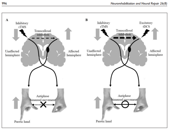
But rTMS has a dark side. It has a negative effect on bimanual movement, decreasing the ability for a person to move both hands. To negate this effect, transcranial direct current stimulation (tDCS) was used. tDCS is not a type of TMS. In fact, tDCS is weaker than TMS and is less prone to cause seizures (to learn more about tDCS, click here). The tDCS was able to counteract the bad side of rTMS, allowing the subjects the ability of bimanual movement.
This picture illustrates the effect of the tDCS against rTMS, as I had stated above. (Picture taken from article)
References:
  1. Takeuchi, N.; Tada, T.; Matsuo, Y.; Ikoma, K.Low-frequency repetitive TMS plus anodal transcranial DCS prevents transient decline in bimanual movement induced by contralesional inhibitory rTMS after stroke. Neurorehabil Neural Repair  201226: 988-999.